JeetVet Endoscopy · Clinical Case Record | Endoscopic Management of Gastric Foreign Body
- Share
- Issue Time
- Oct 1,2025
Summary
Veterinary endoscopy provides a minimally invasive, efficient, and safe solution for diagnosing and treating gastric and intestinal foreign bodies in animals, ensuring faster recovery and better outcomes.
Patient Background
1. Basic Information
Age: 3 months
Species: Blue-and-Gold Macaw
2. Chief Complaint:
Vomiting, poor spirit.
Routine Examination
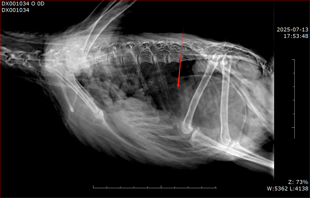
Imaging Evaluation:
X-ray showed a low-density tubular shadow in the proventriculus and ventriculus. Biochemistry revealed no abnormalities.
Diagnosis & Treatment Plan
1. Diagnosis:
Foreign body in the proventriculus and ventriculus.
2. Treatment Outcome:
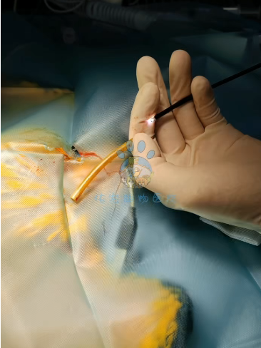
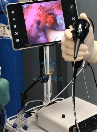
Under inhalation anesthesia with endotracheal intubation, a 3 mm diameter endoscope was inserted through the crop to explore and successfully remove the foreign body.
3. Postoperative Care:
Sucralfate and ampicillin were administered postoperatively to control mucosal damage and secondary infection. Prognosis was favorable.
Analysis & Discussion
The success of this case can be attributed to two key factors:
1. Accurate and Efficient Diagnostic System
Based on the detailed clinical history provided by the owner, combined with imaging results, a rapid and precise diagnosis was achieved, laying a solid foundation for targeted treatment.
2. Significant Advantages of Minimally Invasive Endoscopy