Patient Record: The “Hidden Crisis” of a 4-Month-Old Puppy
- Share
- Issue Time
- Oct 31,2025
Summary
In modern veterinary practice, accurate diagnosis and timely intervention are crucial — especially for young animals. This real case of a 4-month-old puppy named Dundan highlights how a veterinary endoscope played a vital role in detecting and removing a hidden gastric foreign body.
Name: Dundan
Age/Sex: 4 months old, male
Health Background: Fully vaccinated (distemper, parvovirus, etc.), dewormed as scheduled, regular diet.
Reason for Visit: Frequent vomiting (yellow fluid/undigested kibble) and diarrhea (mucus in stool) for three days. Despite good appetite and energy, the owner assumed it was “sensitive digestion” and did not seek help until the symptoms worsened.
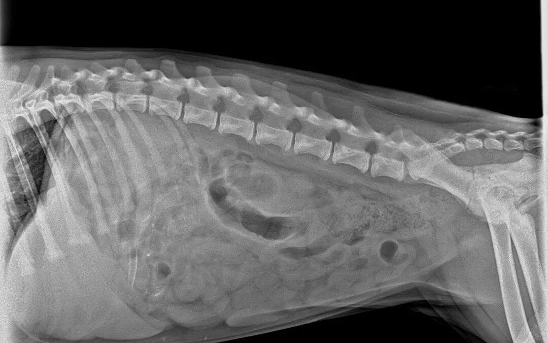
DR Imaging Locked on the Foreign Object
Examination Result: Digital radiography (DR) revealed a high-density linear foreign object in the stomach. Intestinal obstruction was ruled out, and the diagnosis was “gastric foreign body.”
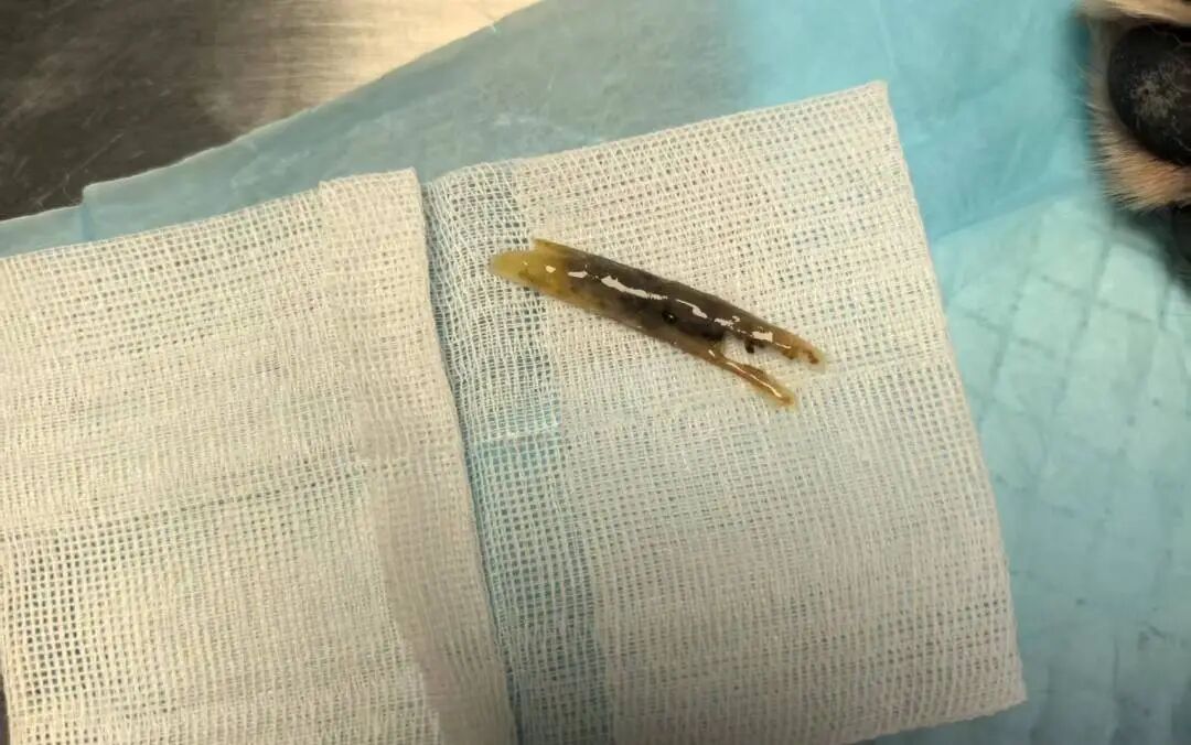
Treatment Plan: Endoscopy was performed to confirm the position and remove the object promptly to prevent blockage. The procedure was done using the JeetVet Veterinary Endoscope.
Treatment Result: The procedure was completed in just 10 minutes. The foreign object was successfully removed, and Dundan recovered well.
Postoperative Care: Routine fluid therapy; small, frequent meals.
Vet’s Reminder — These Symptoms = SOS!
If your pet shows any of the following signs, please seek veterinary help immediately:
- ▪️Frequent vomiting of yellow liquid or undigested food
- ▪️Bloody or mucus-filled stool
- ▪️Lethargy, curled posture, or abdominal pain
Puppies can’t say they’re in pain — “abnormal” means “help me.” Dundan was lucky: timely medical care and professional treatment made all the difference. Attentive care and preventive awareness are the best protection for every pet.
Analysis and Discussion
Two key factors contributed to the successful outcome of this case:
1. Accurate and Efficient Diagnostic System
Based on the pet owner’s detailed symptom description combined with imaging results, a rapid and precise diagnosis was achieved, laying a solid foundation for targeted treatment.
2. Advantages of Minimally Invasive Endoscopic Treatment
Using the JeetVet Veterinary Endoscope provided a clear, high-definition view. Its flexible and slender insertion section minimized mucosal irritation and reduced the risk of complications such as intestinal obstruction.