Veterinary Endoscope Image Quality Guide: What Really Matters Beyond Resolution?
- Share
- Issue Time
- Aug 19,2026
Summary
When choosing a veterinary endoscope, image resolution is often one of the first specifications veterinarians compare.
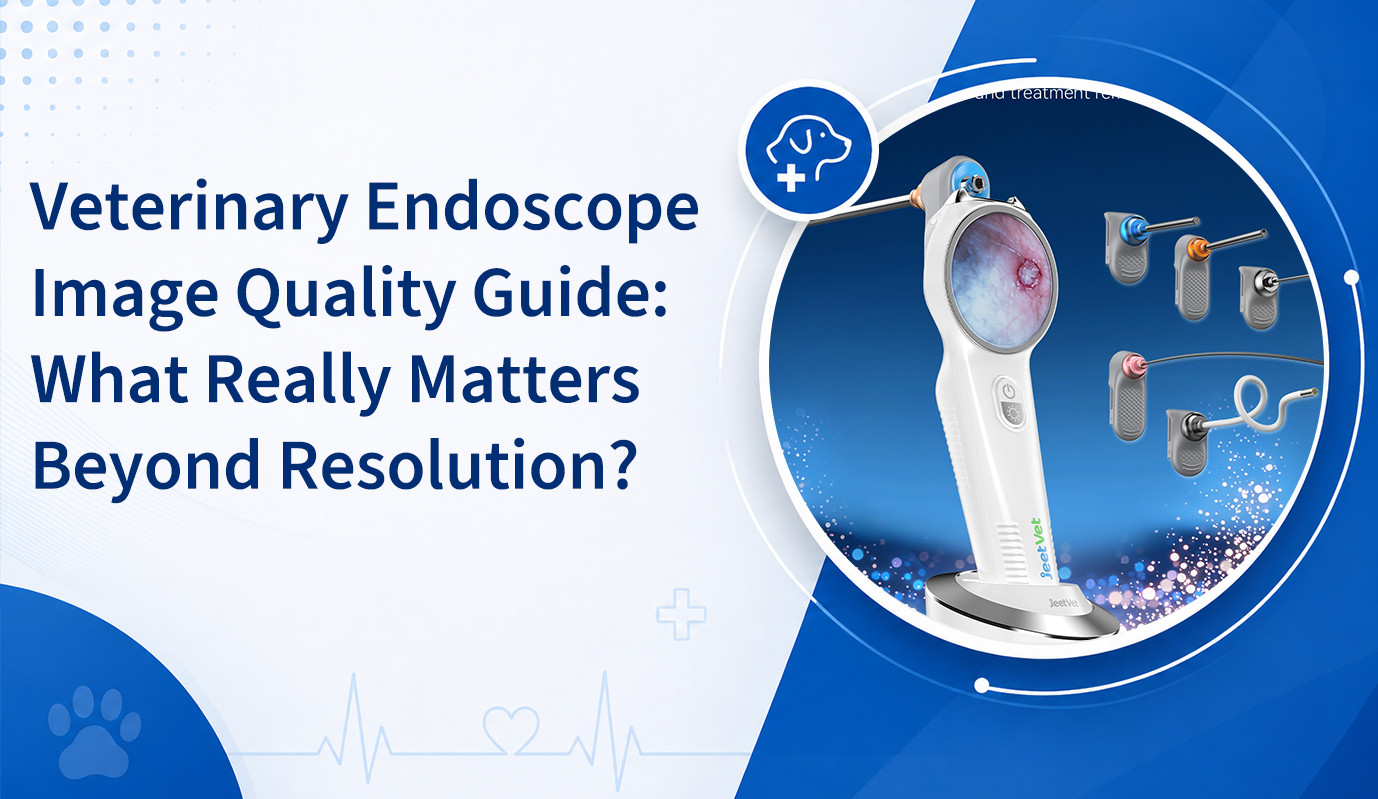
When choosing a veterinary endoscope, image resolution is often one of the first specifications veterinarians compare. High-definition imaging is important, but resolution alone does not determine whether an endoscope can provide clear and clinically useful visualization.
During gastrointestinal, respiratory, nasal, or other endoscopic examinations, veterinarians need to distinguish subtle changes in tissue color, surface texture, bleeding, inflammation, foreign bodies, and abnormal lesions.
To achieve this, several factors must work together—including resolution, illumination, field of view, color reproduction, image stability, focusing performance, and maneuverability.
This guide explains what veterinary clinics should really consider when evaluating veterinary endoscope image quality.
Why Image Quality Matters in Veterinary Endoscopy
A veterinary endoscope provides direct visualization of anatomical structures that may otherwise be difficult to examine. The clinical value of this visualization depends heavily on how clearly the image represents the tissue being observed.
Clear endoscopic imaging may help veterinarians evaluate:
- Mucosal color and texture
- Localized inflammation
- Erosions and ulceration
- Potential bleeding sites
- Foreign bodies
- Abnormal proliferative tissue
- Areas selected for biopsy
Poor visualization can make subtle abnormalities more difficult to identify and may reduce confidence during instrument positioning or tissue sampling.
1. Resolution: Important, but Not the Whole Story
Resolution describes the amount of image detail that an imaging system can reproduce. In general, sufficient resolution can help clinicians observe smaller structures and subtle surface changes more clearly.
However, simply comparing numbers such as 720P or 1080P does not provide a complete picture of real-world endoscopic performance.
Actual image quality can also be affected by:
- Image sensor performance
- Optical design
- Illumination
- Image processing
- Display quality
- Distance between the distal tip and tissue
- Fluid, mucus, blood, or debris in the field of view
For this reason, clinics should evaluate the complete imaging system rather than relying on resolution specifications alone.
2. Illumination: Clear Imaging Starts with Good Light
Internal body cavities are naturally dark. A veterinary endoscope therefore requires reliable illumination to provide useful visualization.
Insufficient lighting may produce dark images and hide important details. Excessive or poorly controlled illumination can create glare and overexposed areas, particularly when the distal tip is positioned close to wet or reflective tissue.
An effective endoscopic imaging system should provide sufficient and relatively even illumination across the observation area.
Good illumination is particularly valuable when examining:
- Gastric mucosa
- Esophageal surfaces
- Nasal passages
- Airway structures
- Small anatomical cavities
3. Color Reproduction: Seeing Tissue as Naturally as Possible
Color can provide clinically useful information during endoscopic examination.
Veterinarians may assess differences in:
- Redness
- Paleness
- Localized bleeding
- Inflammatory changes
- Abnormal tissue appearance
If color reproduction is inaccurate, overly saturated, or inconsistent, subtle tissue differences may become more difficult to interpret.
When evaluating a veterinary endoscope, clinics should therefore consider whether the system provides natural and stable color representation under typical clinical conditions.
4. Field of View: How Much Can You See?
Field of view describes how wide an area can be observed through the endoscope at a given position.
A suitable field of view can help veterinarians maintain anatomical orientation and inspect a broader area without excessive repositioning.
This may be especially useful during:
- Gastroscopy
- Foreign body localization
- Inspection of larger cavities
- Systematic mucosal examination
However, field of view should be evaluated together with image distortion, working distance, and the intended clinical application.
5. Working Distance and Focus
During veterinary endoscopy, the distance between the distal tip and target tissue changes constantly.
The veterinarian may first need a broader overview of an anatomical region and then move closer to inspect a suspicious lesion or position an instrument.
A clinically practical veterinary endoscope should maintain useful image clarity across the working distances required for its intended procedures.
6. Image Stability During Movement
Endoscopy is a dynamic procedure. The insertion tube moves, the distal tip articulates, the patient may have respiratory movement, and instruments may be introduced through the working channel.
For this reason, image quality should not only be evaluated when the endoscope is stationary.
Clinicians should also consider:
- Motion smoothness
- Image latency
- Image stability during articulation
- Clarity during instrument manipulation
A responsive image can make navigation and instrument positioning more intuitive during clinical procedures.
7. Lens Cleanliness and Fluid Management
Even an advanced imaging system cannot provide a clear image if the distal lens is covered by mucus, blood, food residue, or other material.
Depending on the endoscope design and procedure, effective air, water, suction, or flushing capabilities can help maintain a usable visual field.
This is particularly important during gastrointestinal procedures where secretions and debris may interfere with observation.
What Should Clinics Evaluate Before Purchasing?
Instead of comparing only resolution numbers, veterinary clinics should assess image performance as part of the entire endoscopy system.
| Factor | What to Evaluate |
|---|---|
| Resolution | Whether clinically relevant details are clearly visible |
| Illumination | Brightness, uniformity, and control of glare |
| Color | Natural and stable tissue representation |
| Field of View | Coverage appropriate for intended procedures |
| Working Distance | Useful clarity at typical examination distances |
| Motion Performance | Smooth visualization during navigation and articulation |
| Documentation | Ability to capture and review images or video when required |
Image Quality and Endoscopic Biopsy
Image quality becomes particularly important when a veterinary endoscope is used for targeted biopsy.
The veterinarian must first identify an appropriate sampling location and then position biopsy forceps accurately under direct visualization.
Clear imaging can help maintain orientation while the instrument enters the field of view and approaches the target tissue.
This illustrates why image quality, maneuverability, and working channel performance should be considered together rather than as separate specifications.
How JeetVet Approaches Veterinary Endoscope Imaging
JeetVet develops veterinary endoscope solutions for companion animal and exotic animal applications, with a focus on combining practical imaging performance with maneuverability and clinical usability.
Depending on the model and intended application, JeetVet veterinary endoscopy solutions are designed to support clear visualization for procedures such as:
- Gastrointestinal examination
- Foreign body localization and retrieval
- Targeted biopsy
- Respiratory examination
- Selected exotic animal applications
For veterinary practices, the goal should not simply be to select the highest resolution number. A practical endoscopy system should provide a balanced combination of clear imaging, reliable illumination, flexible navigation, appropriate working channel capability, and convenient clinical operation.
Conclusion
Image quality is one of the most important considerations when choosing a veterinary endoscope, but resolution tells only part of the story.
Illumination, color reproduction, field of view, working distance, motion performance, lens cleanliness, and image stability all influence what the veterinarian actually sees during a procedure.
By evaluating the complete imaging system rather than focusing on a single specification, veterinary clinics can select an endoscope that better matches their patients, procedures, and daily clinical workflow.