How Does the Veterinary Endoscope Work?
- Share
- Issue Time
- Mar 13,2025
Summary
Learn about the working principle of veterinary endoscopes and why they are essential in modern animal healthcare.
A portable veterinary endoscope is a specialized medical device used to examine the internal organs and structures of animals. It consists of a long, flexible or rigid tube equipped with a camera and light source, allowing veterinarians to visualize areas such as the gastrointestinal tract, respiratory system, and urinary tract without the need for invasive surgery. In this article, JeetVet will guide you on the work principle of veterinary endoscope, and explore the technology behind veterinary endoscope, their key components, and how they are transforming veterinary practices worldwide.
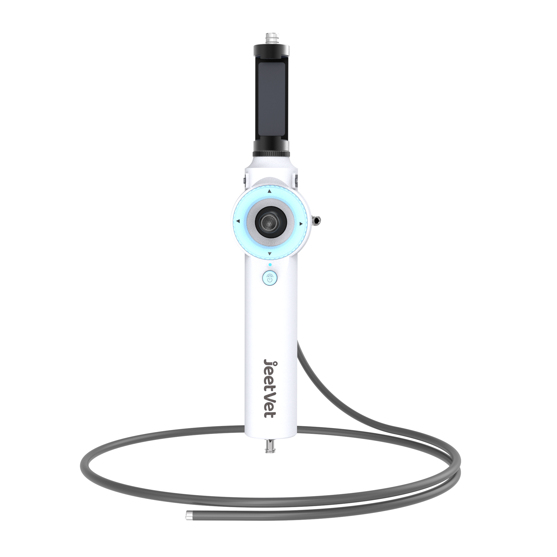
Key Components of a Veterinary Endoscope
Understanding the technology behind portable veterinary endoscope starts with its core components:
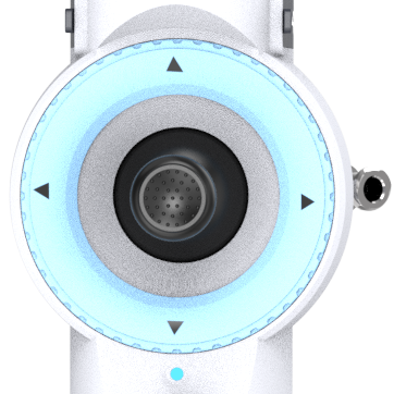
1. Camera
The camera is the most critical component of a veterinary endoscope, providing high-resolution images for accurate diagnosis. Modern endoscopes often feature 4K imaging technology, which offers exceptional clarity and detail.
Key Features:
High-Resolution Imaging: Captures detailed images of internal structures
Zoom Capability: Allows for closer examination of specific areas
Low-Light Performance: Ensures clear imaging in dark cavities
2. Light Source
The light source provides illumination for the camera, ensuring that the internal structures are clearly visible.
Key Features:
Brightness: Adjustable light intensity for optimal visibility
Consistency: Stable illumination without flickering
Durability: Long-lasting and energy-efficient
3. Insertion Tube
The insertion tube is the part of the endoscope that is inserted into the animal's body. It can be either flexible or rigid, depending on the application.
Key Features:
Flexibility: Allows navigation through complex pathways
Durability: Resistant to bending and breaking
Diameter: Varies depending on the size of the animal and the area being examined
4. Control System
The control system allows the veterinarian to maneuver the endoscope with precision. It typically includes knobs or levers for controlling the movement of the insertion tube.
Key Features:
Precision: Fine-tuned controls for accurate navigation
Ergonomics: Designed for comfortable use during prolonged procedures
Responsiveness: Immediate feedback for smooth operation
5. Working Channel
The working channel is a small passage within the endoscope that allows specialized tools to be passed through. These tools can be used for various therapeutic procedures, such as biopsies or foreign body removal.
Key Features:
Versatility: Compatible with a range of tools and accessories
Size: Varies depending on the intended use
Accessibility: Easy to clean and maintain

Working Principle of the Veterinary Endoscope
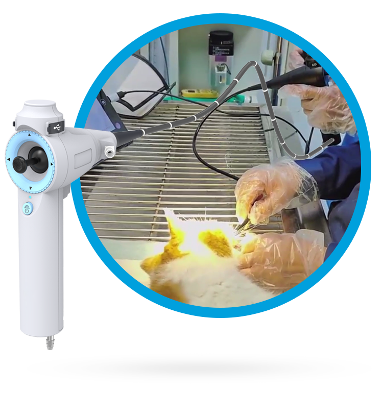
The animal endoscope combines advanced imaging and fiber-optic technology to provide real-time visualization of internal structures. Here's a step-by-step breakdown of how it works:
Step 1: Insertion
The endoscope is gently inserted into the animal’s body through a natural opening (e.g., mouth, nose) or a small incision.
Step 2: Imaging
The camera at the tip of the endoscope captures high-definition images, which are transmitted to a monitor in real-time.
Step 3: Navigation
The veterinarian uses controls to maneuver the endoscope, allowing them to explore hard-to-reach areas with precision.
Step 4: Intervention
If necessary, specialized tools can be passed through the endoscope to perform procedures like biopsies, polyp removal, or stent placement.
Conclusion
Understanding the working principle of portable veterinary endoscope is essential for maximizing its potential in modern veterinary care. From high-resolution imaging to precise control, each component plays a crucial role in ensuring accurate diagnostics and effective treatments. Ready to explore the benefits of veterinary endoscope for your practice? We offer specific endoscopy solutions for small animals, large animals, exotic pets, veterinary clinics, and pet hospitals, visit to learn more!


